Dr. Kämpf, Prof. Dr. Ketelsen, Dr. Trübenbach and their team of highly qualified physicians regularly participate in the external quality assurance for curative mammography of the “Kassenärztliche Vereinigung” in order to offer you the highest quality.
When is breast imaging indicated?
Breast imaging is recommended in the following situations:
- Palpable lumps or areas of induration
- Breast pain or structural changes
- Nipple discharge
- Suspicious or unclear prior findings
- Follow-up after breast cancer treatment
The aim of imaging is to reliably differentiate between benign and malignant changes and to guide further diagnostic or therapeutic steps.
Our Diagnostic Modalities
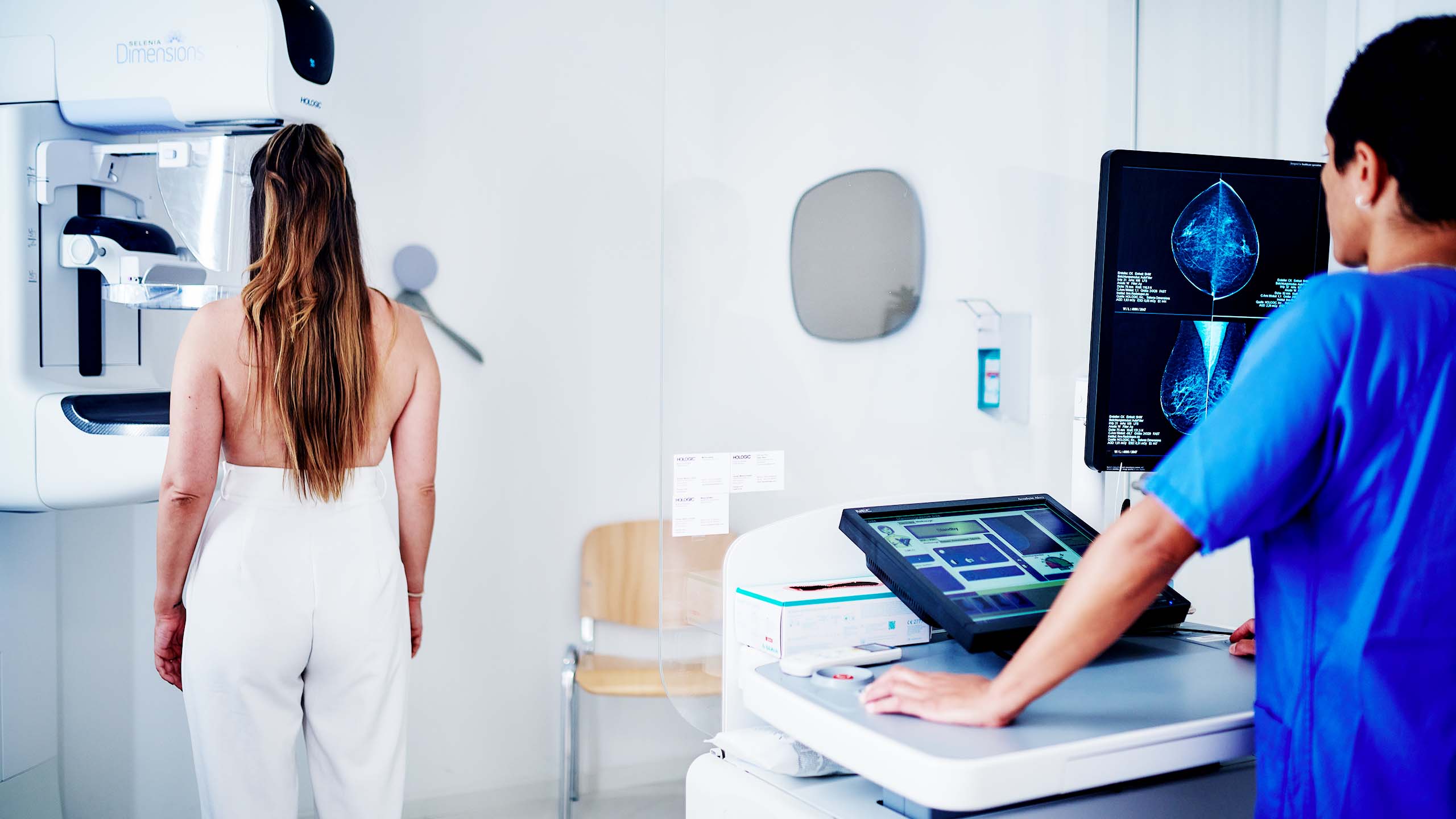
Mammography
Mammography is the primary imaging modality for detecting microcalcifications and structural abnormalities of the breast. It plays a central role in the curative evaluation of suspicious findings.
Ultrasound (Sonography)
Ultrasound complements mammography and is particularly useful for evaluating masses, cysts, and dense breast tissue. It allows for real-time, detailed assessment and often contributes directly to clinical decision-making.
Individualized and Guideline-Based Assessment
The combination of imaging modalities enables a precise and individualized diagnostic approach.
All examinations are performed in accordance with established guidelines and standardized classification systems (e.g., BI-RADS), ensuring consistent and transparent reporting.
Interdisciplinary Collaboration
Breast imaging is carried out in close collaboration with gynecologists, oncologists, and surgeons.
Our reports provide clinically relevant information that supports further treatment planning and decision-making.
Advanced Technology and Expertise
Our technical infrastructure and specialization ensure:
- High-resolution mammography
- Modern ultrasound diagnostics
- Fast and reliable evaluation of even complex cases
Your Benefits
- Specialized breast imaging in a curative setting
- Combination of established imaging modalities
- Standardized reporting using BI-RADS
- Close collaboration with referring physicians
- State-of-the-art technology and rapid appointment availability