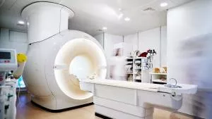
Modern cardiac imaging - CT and MRI diagnostics for your heart.
One focus of our team is cardiodiagnostics. Our specialist team of radiologists and cardiologists bundles a maximum of expertise and clinical experience for you. We offer both cardiac MRI and cardiac CT at various locations. Please feel free to call us for a consultation.
Our specialists Prof. Dr. Fenchel and Prof. Dr. Ketelsen are certified cardiovascular radiologists of the German Radiological Society (DRG) and work closely together with our specialist for internal medicine and cardiology, Dr. Nico Merkle, in order to combine a maximum of expert knowledge and clinical experience. Our imaging center in Mitte is certified as a Center for Cardiovascular Imaging by the German Radiological Society.
In our radiological care centers we offer both cardiac MRI and cardiac CT.
Note: The cost of cardiac MRI and cardiac CT are usually covered by the private health insurance companies. Statutory health insurance only covers the costs in certain cases.
To advise you, we have set up a special heart hotline for you.
We are at your disposal under 030 322 913 207 or by using our contact form.
Cardiac MRI.
Cardiac MRI is a high-performance tool, with the flexibility to evaluate a wide array of cardiac pathology. That tremendous flexibility, however, entails that each examination must be specifically tailored to answer the precise clinical questions that are posed. There is no single “standard” cardiac MRI examination. For this reason, it is imperative that when requesting a cardiac MRI, enough clinical history is provided to allow the study to be specifically tailored to address all of the clinically-relevant questions.
In general, the examination consists of two parts: pre-contrast series, and post-contrast series. The pre-contrast images are designed to evaluate cardiac anatomy, function, and quantitative flow. Post-contrast images consist of several different types:
- Perfusion images, which are images of the myocardium obtained as the contrast is injected intravenously.
- Delayed enhancement images, which are usually obtained 10-15 minutes after contrast injection, to assess for abnormal gadolinium contrast enhancement within the myocardium (often called “late gadolinium enhancement”, or “LGE”). These images provide information on myocardial scarring or fibrosis due to both ischemic and non-ischemic causes.
Cardiac MRI requires some preparation. ECG electrodes will be placed on the chest to allow for cardiac motion gating. Because respiratory motion can significantly degrade image quality, patients will be asked to hold their breath repeatedly during image acquisitions, usually between 10-20 second per breath hold. The examination itself can take between 30 and 60 minutes, and patients must be able to lie flat and still for this amount of time.
Cardiac-CT.
Calcium Score CT:
This examination requires the placement of ECG electrodes on the chest. The CT scanner uses ECG-tracking or “gating” to minimize cardiac motion and provide unenhanced (i.e. no IV contrast used) CT images of the heart. These images are analyzed to provide a quantitative evaluation of coronary artery calcification (i.e. “Calcium Score” or “Agatston Score”).
The CT Calcium Score is currently recommended as a stand-alone test for asymptomatic patients because the CT Calcium Score correlates with future risk of adverse cardiovascular events (e.g. myocardial infarction). Current national and international guidelines recommend Calcium Score CT as a risk stratification tool in patients that are at intermediate risk for cardiovascular disease (e.g. based on Framingham risk factors).
Coronary CT Angiography:
Our standard cardiac CT examination has two parts. First, we begin with an unenhanced CT of the heart for CT Calcium Scoring (as described above). Before proceeding to the second part of the exam, these images are immediately analyzed. If a patient has a large volume of coronary artery calcification, further assessment with CT angiography may be precluded because in the presence of extensive calcification evaluation of the coronary artery lumen is significantly impaired.
If appropriate, the second part of the CT examination, the cardiac CT angiogram, is performed. To optimize imaging, the patient’s heart rate should be below 65 BPM. To achieve this rate, oral and/or intravenous beta-blocker will be administered in the radiology department one hour to several minutes prior to the examination (if no contraindications exist). An 18- or 20-gauge venous catheter is placed into a cubital vein for administration of non-ionic intravenous contrast agent. If appropriate, a dose of sublingual nitroglycerine will also usually be given to the patient immediately prior to image acquisition for the cardiac CT to help dilate the coronary arteries. Both parts of the study require the patient to hold their breath for approximately 10-20 seconds (the time it takes to completely scan the heart). The entire CT examination takes approximately 15-20 minutes. Patients may return to normal activity immediately following the examination.
Contraindications.
Contraindications to cardiac CT angiography include cardiac arrhythmias and renal impairment. Prior contrast reactions are a relative contraindication; such patients can frequently be pre-treated with the use of a steroid and anti-histamine preparation.
Target Patient Population
For Calcium Scoring CT:
- Asymptomatic patients with recognized cardiac risk factors (e.g. hypertension, hyperlipidemia, smoking history, or a family history of heart disease)
For Coronary CT Angiography:
- Symptomatic patients with chest pain, equivocal clinical presentation or indeterminate prior non-invasive cardiac tests
- Patients with cardiac vascular anomalies
- Patients with bypass grafts
- Patients with congenital heart disease (e.g. to assess cardiac and vascular anatomy)
- Patients with potential pulmonary venous abnormalities (e.g. post pulmonary vein ablation/isolation to assess for pulmonary vein stenosis)
Our state-of-the-art CT scanner (including 128-slice computed tomography) allows us to image the heart with the highest temporal and spatial resolution at the lowest possible radiation exposure. If clinical indication for imaging is correct (see above), cardiac CT can save unnecessary cardiac catheter examinations.


